Study protein functional states and interplay - Unique Product!
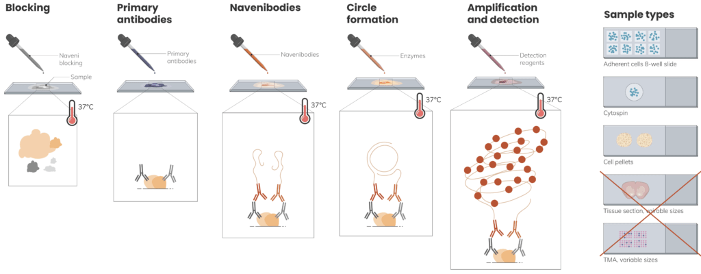
Naveni™TriFlex Cell is designed for fluorescence detection of two proteins in a free and complex state. Based on the Naveni™ Proximity Ligation Technology, the in situ kit provides accurate data with precise staining of total protein A, total protein B, and the interaction AB between the two proteins.
Validated for cell samples
To be used with an own primary mouse and rabbit antibody pair
Very easy to perform (identically to an immunostaining)
Compatible with regular immunostaining equipment (FITC, Cy3, Cy5 filters)
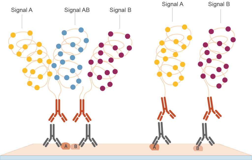
Naveni™ TriFlexrelies on two user-determined primary antibodies against the targets of interest, and on proprietary TriFlexNavenibodies (in orange), which are antibody-based proximity reagents. Only proteins located at <40 nm distance are recorded as interacting. Naveni™ TriFlexdetects total protein A (i.e., both free and in complex with B, yellow), total protein B (maroon), and the AB interaction (blue). The detected A, B and AB signals are amplified and generate fluorescent readout in three channels corresponding to each protein pool.
Naveni™ TriFlex Application examples
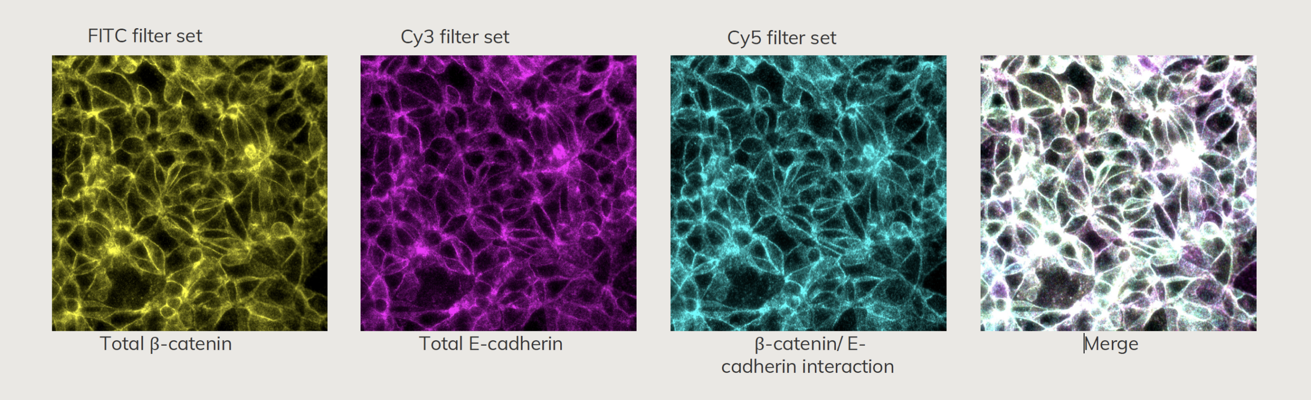
Naveni™ TriFlex Cell - Protein interaction
β-catenin/ E-cadherin staining in MCF7 cells, 40x objective. β -catenin and E-cadherin are highly expressed proteins and the interaction between the two proteins is important for cell-cell adhesion.
Naveni™ TriFlex Cell - From Birth to Death
NaveniTMTriFlex Cell was used to simultaneously detect free and interacting Lamin B1 and Histone H3, 40x objective, deconvolved.
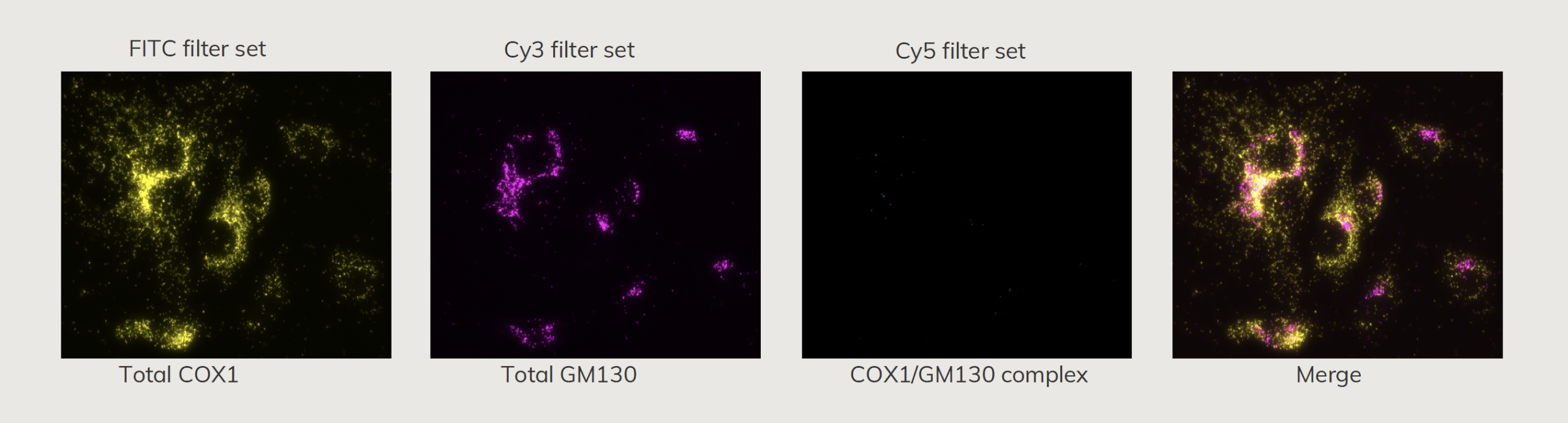
Naveni™ TriFlex Cell - Co-localization is not a synonym to protein interaction
NaveniTMTriFlex Cell was used to simultaneously detect total COX1 and GM130, and their potential interaction. This assay was used as a biological negative control, as COX1 and GM130 belong to different cell compartments and thus do not interact, what is visualized using the Cy5 channel. Note that in the merged image, however, COX1 and GM130 appear partially co-localizing (white) where signals are particularly dense.